Prosopometamorphopsia: The Brain that blurs faces
In some rare conditions, faces remain recognizable but appear distorted. These alterations provide new insights into how perception works.
His name is A.D., 59 years old. A right-handed man with no remarkable medical history, fully autonomous, no psychiatric scars or developmental concerns. Just an ordinary individual until one day, something quietly unraveled behind the curtain of appearances. Since a minor stroke, faces have changed in nature. Every time he sees one, a strange asymmetry emerges. The right half appears intact, familiar. The left side, however, is deformed melting, unstable, as though wax had run down under the sun. This isn’t a fleeting illusion. It’s not a passing glitch. It’s a lasting fracture in his ability to perceive others.
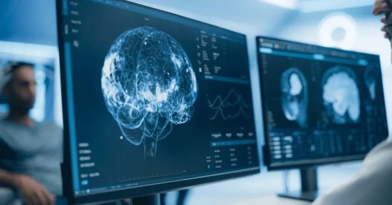
Still, A.D. recognizes people. He knows who they are, can name their voices, and recalls their stories. But he no longer sees a face as a unified whole. One half is clear; the other slips away like a shattered reflection. Brain imaging reveals the origin of the disruption: a lesion in the left splenium of the corpus callosum, the neural bridge between both hemispheres. A region where visual input becomes integrated where fragments join to form a complete face. That is precisely where the fault line lies.
This case, reported by Jorge Almeida and colleagues in Current Biology (2020), illustrates a rare and puzzling visual disturbance: prosopometamorphopsia. Unlike prosopagnosia, where facial identity is lost, here identity is preserved. It’s the structure that collapses. Facial features warp, liquefy, dissolve. You know who you’re looking at but the face doesn’t hold together.
🔗Explore further: How the human brain works
The brain’s invisible blueprint
Seeing a face isn’t simply about capturing an image on the retina. It’s a complex perceptual act, carried out in milliseconds by a highly specialized chain of neural processes. The human face is one of the richest and most vital visual stimuli in our environment. It conveys identity, emotion, intention, and nonverbal communication. That’s why the brain begins to focus on faces from the earliest days of life. Newborns instinctively turn toward facial configurations, even basic schematic ones.
🔗 Read also: When the world distorts: The neuroscience behind Alice in wonderland syndrome
This early preference is accompanied by the gradual development of a dedicated neural system. Central to this network is the Fusiform Face Area (FFA), a region located in the lateral-inferior fusiform gyrus within the occipitotemporal cortex. The FFA is selectively activated during facial perception far more than when viewing objects, scenes, or words. But face processing goes beyond the FFA. It involves the Occipital Face Area (OFA) for analyzing facial parts (eyes, nose, mouth), and parietal regions responsible for spatial integration and attentional orientation.
Together, these areas deconstruct the face into perceptual units, analyze the spatial relationships between features, and reassemble them into a coherent whole. This process depends on continuous comparison between visual input and an internal facial prototype a mental template refined through experience. This internal model helps us recognize faces despite changes in perspective, lighting, expression, or movement.
Perceiving a face, then, means more than spotting individual features; it involves organizing them into an expected structure. This demands close cooperation between both hemispheres. When any part of this chain is disrupted whether in local detection, spatial integration, or internal comparison the entire perception may become unstable or unrecognizable, as in A.D.’s case.
His lesion in the left splenium of the corpus callosum critical for interhemispheric communication appears to impair this integration. The brain still receives visual data but fails to merge it into a stable form. Recognition persists, but proper facial construction breaks down. The issue is not memory, but perceptual coherence. The features are present, but distorted, displaced, structurally incoherent. Perception becomes unstable, often unsettling, and resists conscious correction.
This case highlights the visual system’s complexity. Facial perception depends on a fragile balance between sensory input, internal representations, and hemispheric coordination. When this balance is disturbed, facial memory remains but visual stability collapses. The issue isn’t who the person is, but how their face is perceived. The once-familiar face becomes unstable, distorted, foreign. And within this perceptual gap, the deep-rooted feeling of familiarity the silent foundation of social connection begins to crumble.
🔗 Explore further: When familiar faces become strangers: Unraveling Capgras syndrome
When the brain fills in the blanks
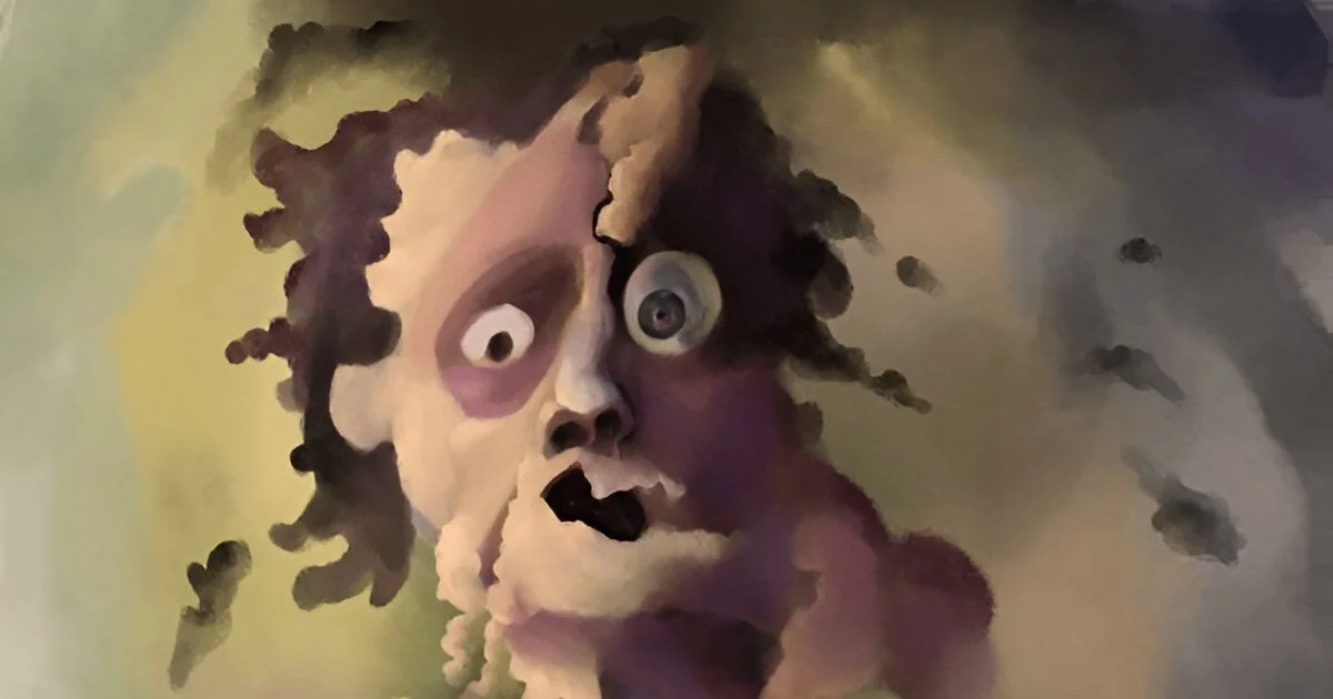
A major breakthrough came in 2024 with a study by Mello et al., published in The Lancet. For the first time, researchers managed to visually recreate how a person with prosopometamorphopsia actually sees faces. The patient described distorted faces with elongated features, some even appearing “demonic.” Curiously, these distortions only occurred during live, face-to-face interactions and vanished completely when viewing photos.
Brain imaging revealed another lesion, this time in the head of the left hippocampus. While the hippocampus is widely known for memory processing, it also plays a role in contextual evaluation and the sense of familiarity. The link between live perception, relational context, and visual distortion suggests that face perception involves more than structure it depends on the situation in which the face appears.
To objectively capture the patient’s experience, researchers used an AI-driven image transformation tool to generate photorealistic visuals based on his detailed descriptions. The resulting images closely matched what he claimed to see. This methodology ushers in a new era in the study of perceptual disorders where subjective experience can become partially visualized and empirically explored.
The findings confirm that prosopometamorphopsia is not just a local glitch in visual processing. It is not a defect in a single brain module or peripheral structure but rather a broader network dysfunction affecting various layers of processing.
🔗 Discover more: The face that disappears: Inside the mind with prosopagnosia
This typically resilient system allows us to recognize faces even when they’re partially obscured, poorly lit, or distorted by emotion. However, such flexibility depends on finely tuned balance. A small lesion whether in the corpus callosum, hippocampus, or fusiform gyrus can throw the entire network off sync, disrupting coordination between perceptual, mnemonic, and emotional components. The face remains recognizable, but its form becomes unstable, unsettling, even frightening.
Facial recognition, then, is never a passive act. It is a dynamic reconstruction, built moment by moment from shifting cues, stabilized by a brain constantly predicting, adjusting, and reassembling. When this process breaks down, it’s not just the perception of others that falters it’s the sense of security and connection that a familiar face represents. In this way, prosopometamorphopsia is more than a symptom it reveals the silent complexity behind our ability to truly see another human being.
Références
Almeida, J., Freixo, A., Tábua-Pereira, M., Herald, S. B., Valério, D., Schu, G., … & Duchaine, B. (2020). Face-specific perceptual distortions reveal a view- and orientation-independent face template. Current Biology, 30(19), 4071–4077.
Mello, A., Stehr, D., Bujarski, K., & Duchaine, B. (2024). Visualising facial distortions in prosopometamorphopsia. The Lancet, 403(10391), 1176.
Sara Lakehayli
PhD, Clinical Neuroscience & Mental Health
Associate member of the Laboratory for Nervous System Diseases, Neurosensory Disorders, and Disability, Faculty of Medicine and Pharmacy of Casablanca
Professor, Higher School of Psychology