How the human brain works
The human brain is the most complex organ we know. How can a few hundred grams of tissue interpret the world, learn from experience, and constantly adapt? This guide explores the neuroscience behind thought, memory, and perception.
Understanding how the human brain works is not simply a matter of describing a biological organ. It involves grasping the logic of a system capable of perceiving, anticipating, learning, and continually transforming itself. With around 86 billion neurons interconnected by hundreds of trillions of synapses, the brain is one of the most complex organized systems known to date. Its complexity, however, does not lie only in the number of its cells. It resides above all in the dynamics of their interactions. The brain is not a fixed structure. It is a living network in continuous activity whose organization evolves with every experience.
Contemporary neuroscience shows that brain activity rests on a dual reality. On the one hand, there is a specialized anatomical architecture composed of differentiated regions and functional circuits. On the other hand, there is a permanent capacity for adaptation that allows these circuits to change depending on learning, context, and time. This tension between stability and transformation is one of the fundamental principles of brain function.
Long perceived as a hierarchical command center, the brain is now understood as a distributed system. Cognitive functions are not localized in a single point. They emerge from the coordinated interaction of neuronal networks spread across different regions. Memory, attention, perception, and language do not correspond to isolated compartments but to dynamic patterns of activity that are continuously modulated by context and experience.
This perspective profoundly transforms how we approach the human brain. Studying it does not mean reducing human beings to a set of circuits. Rather, it means illuminating the biological conditions that make thought, emotion, and memory possible while acknowledging the irreducible complexity of the living system that supports them.
This guide explores the fundamental mechanisms that structure this organization in order to better understand how biological tissue can generate a coherent experience of the world and of the self.
The brain as a networked organ
Contrary to the traditional image of a single command center, the brain does not rely on one conductor directing the orchestra. Instead, it functions through a vast ensemble of interconnected networks whose coordination supports all mental activity.
These networks consist of populations of neurons distributed across various regions of the cortex, brainstem, and limbic system. A cognitive function such as decision making or planning does not appear in an isolated box within the brain. It emerges from the coordinated interaction of multiple regions working in parallel.
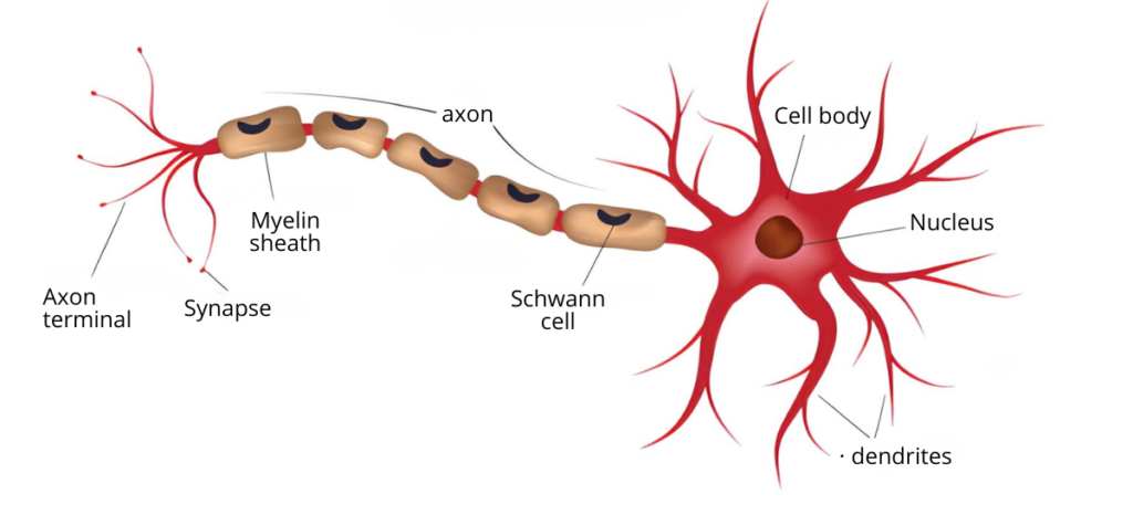
Emergence: At the microscopic level, each neuron generates an electrical signal known as an action potential. This signal travels along the axon and reaches the synapses, contact zones where it triggers the release of chemical messengers called neurotransmitters. These molecules modulate the activity of target neurons by increasing or inhibiting their excitability.
Through this electrochemical dynamic, the brain can integrate, transform, and transmit immense quantities of sensory, emotional, and cognitive information. However, this process is not merely an automatic mechanism. It follows organizational principles that favor energy efficiency, rapid processing, and resilience to disturbance.
Unlike a hard drive that passively stores data, the brain constantly reorganizes itself. It continually reshapes its connections in order to maintain the balance and coherence of the neuronal network.
🔗Read also: Brain photons: Discovery of a natural light emission
For a long time, these dynamics were difficult to observe directly. Advances in functional magnetic resonance imaging have made it possible to visualize brain activity in the living human brain. This technique does not measure the electrical activity of neurons directly. Instead, it detects local variations in blood flow associated with their activation.
When the brain engages in a task such as memory or attention, certain regions display an increase in the BOLD signal, which reflects greater oxygen consumption. Analysis of these variations has shown that cognitive functions do not rely on a single area but on networks of interconnected regions that activate in a coordinated manner.
For example, during autobiographical memory tasks, the hippocampus, several temporal regions, and prefrontal areas become active together. Such observations have profoundly reshaped our understanding of brain organization by revealing that cognition emerges from dynamic cooperation between multiple brain regions.
The Main Lobes of the Brain: The human brain is organized into several specialized regions known as cerebral lobes, each associated with particular functions. Although these regions work together in networks, they play distinct roles in perception, thought, and action.
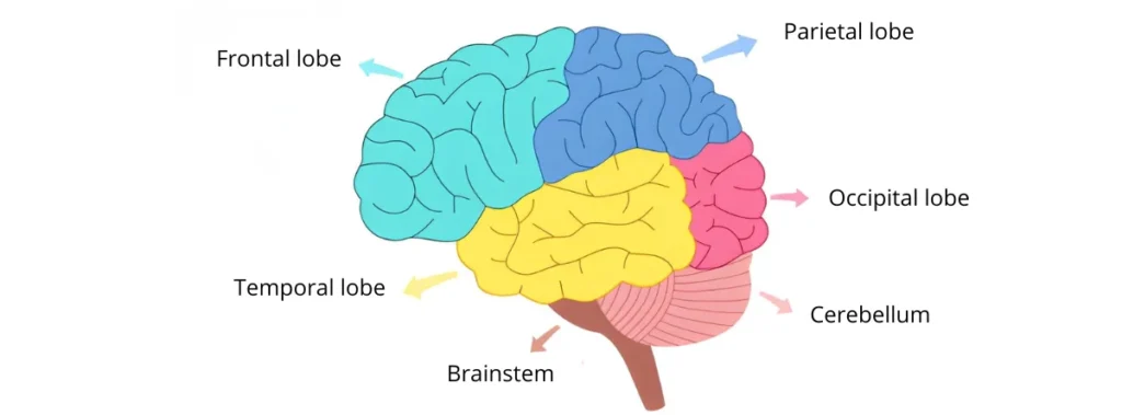
Frontal lobe: Involved in planning, decision making, behavioral control, and voluntary movement.
Parietal lobe: Processes sensory information related to touch, body position, and spatial orientation.
Temporal lobe: Plays a role in hearing, memory, and language comprehension.
Occipital lobe: Specialized in processing visual information.
Brain plasticity: how experience reshapes neural connections
If the brain operates through networks, these networks are not fixed. Their organization evolves continuously. This capacity for adaptation is known as brain plasticity.
Plasticity does not mean that the brain can modify anything at any time. More precisely, it refers to the ability of neuronal circuits to adjust their efficiency and organization in response to experience. This phenomenon relies on identifiable biological mechanisms that have been studied for several decades.
At the microscopic level, plasticity often appears as changes in the strength of synapses. When two neurons are repeatedly and coordinately activated, transmission between them may become more efficient. This process, known as long term potentiation, increases the likelihood that they will activate together in the future. Conversely, some connections may weaken when their activity decreases. This phenomenon is known as long term depression. Such adjustments contribute to the formation and stabilization of learning.
What is brain plasticity? Brain plasticity refers to the capacity of the brain to modify its neuronal connections in response to experience, learning, or injury. Through this mechanism, neuronal circuits gradually reorganize and adapt their functioning to environmental demands.
Plasticity is not limited to local synaptic changes. It can also involve the reorganization of broader neuronal circuits. After brain injury, certain functions may be partially taken over by adjacent or alternative networks. Similarly, during intensive training, some neural pathways gradually become more efficient and better coordinated. Over time, the brain tends to optimize its activation pathways.
Several empirical observations illustrate this phenomenon. Studies conducted with professional musicians have shown increased volume in certain motor and auditory regions compared with non musicians. Among London taxi drivers, researchers have observed enlargement in specific portions of the hippocampus associated with intensive spatial navigation learning. These results suggest that repeated use of a neuronal network can modify its structural organization over the long term. However, such transformations do not mean that the brain can freely reconfigure itself without constraints. They reflect a gradual capacity for adaptation shaped by repeated experience and environmental demands.
🔗Explore further: How Astrocytes Reshape the Brain
This capacity also varies across the lifespan. During childhood and adolescence, certain developmental windows favor the rapid acquisition of abilities such as language or specific sensory skills. In adulthood, plasticity remains present but operates within a more constrained framework. Stabilized circuits provide strong functional robustness, though with less flexibility than during early development.
Plasticity therefore always operates within biological limits. It depends on energetic, structural, and temporal conditions that frame any lasting modification of circuits. Transforming a network is not simply a functional decision. It involves molecular, cellular, and sometimes anatomical adjustments regulated by the physiology of the system itself. The idea of a brain capable of unlimited transformation is appealing but inconsistent with current scientific evidence. Adaptation is real, but it unfolds within a precise physiological framework.
This tension between stability and transformation is fundamental. Without stability, memory could not persist. Without plasticity, learning would not be possible. Brain function relies on this delicate balance.
Three levels of plasticity: Synaptic plasticity refers to changes in the efficiency of connections between neurons.
Functional plasticity refers to the reorganization of networks involved in a given task.
Structural plasticity refers to more durable changes such as dendritic growth, myelin modification, or broader circuit reorganization.
How memory works in the brain
If plasticity allows the brain to modify its connections, memory corresponds to the relative stabilization of these modifications over time. Memory is not a compartment where information is stored. It is a dynamic process through which certain patterns of activity become more probable, more durable, and more easily reactivated.
Remembering therefore does not mean preserving an intact record of the past. It involves transforming the organization of a neuronal network. Each memory corresponds to a modification in the connections and activity dynamics that structure the brain.
The formation of a memory unfolds through several stages. The first stage is encoding, when an experience temporarily modifies the activity of certain neuronal circuits. At this stage the trace remains fragile. Its persistence depends on factors such as attention, repetition of the experience, and the emotional context in which it occurs.
Next comes consolidation, which gradually stabilizes this trace. This process relies on durable synaptic modifications and a redistribution of activity across different brain regions. The hippocampus plays a central role in this initial phase by coordinating information from multiple cortical areas. Over time, some memory traces become less dependent on the hippocampus and become more widely embedded within cortical networks.
🔗Discover more: The eternal memory: When love defies forgetting
Sleep and memory consolidation: why the brain needs sleep to learn
Sleep plays an active role in the consolidation of memories. During certain phases of slow wave sleep and rapid eye movement sleep, coordinated reactivations of the networks involved in recent learning have been observed. These reactivations are thought to strengthen relevant connections and integrate new information with knowledge that has already been established.
Electrophysiological recordings have shown that during deep slow wave sleep, sequences of neuronal activity originally present during learning are partially replayed. This phenomenon, known as reactivation or replay, is believed to promote the strengthening of certain synaptic connections and the gradual stabilization of memory traces.
Conversely, experimental studies indicate that sleep deprivation occurring after learning can impair consolidation, particularly for certain forms of declarative memory. Sleep therefore appears not as a passive state, but as an active phase of reorganization and integration of lived experiences.
Sleep and the Replay Phenomenon: Studies conducted in animals have shown that during deep slow wave sleep, sequences of neuronal activity observed during learning can be partially replayed. This phenomenon, known as hippocampal replay, has been described in particular in the work of György Buzsáki and his collaborators on hippocampal oscillations. In rats, intracerebral recordings have revealed that neurons activated during spatial navigation tasks reactivate during subsequent sleep, often on a compressed timescale. These reactivations are accompanied by brief synchronized bursts of activity known as sharp wave ripples, which are considered a key mechanism in the communication between the hippocampus and the cortex. Replay therefore does not correspond to a faithful “movie” replaying in the brain. Instead, it reflects a partial and selective reactivation of neuronal networks involved in recent learning. This reactivation is thought to strengthen certain synaptic connections and help integrate new information with knowledge that has already been established.
🔗Read also: Sleepless Nights: How sleep deprivation strengthens unwanted memories
How the brain stores memories: a distributed and dynamic system
Remembering does not involve retrieving a perfectly preserved fragment of the past. It involves reactivating networks whose activity has been progressively shaped by experience.
No memory resides in a single cell or in a precise point of the brain. Neuroimaging and lesion studies show that memory traces are distributed across multiple interconnected regions.
The different components of a single experience visual, auditory, emotional, contextual engage distinct areas. Structures such as the hippocampus coordinate these elements by linking them into a coherent configuration.
This distributed organization explains both the resilience and the vulnerability of memory. A localized lesion may alter one aspect of a memory without erasing it entirely, whereas degenerative diseases may gradually disrupt the networks responsible for stabilizing memories.
Memory is not only distributed. It is also dynamic. Each recall temporarily destabilizes the memory trace before it becomes stabilized again in a process known as reconsolidation.
Remembering therefore does not mean replaying a fixed recording. It means reconstructing an event from neuronal fragments and current knowledge. Memories evolve, reorganize, and adapt over time.
🔗Explore further: False memories: The brain’s illusion of truth
How the brain anticipates the world
Describing the brain as a plastic and organized network does not fully capture its deeper logic. Contemporary research suggests that the brain does not merely react to the world. It constantly anticipates it.
According to several current theoretical models, brain activity relies on mechanisms of continuous prediction.
The brain constantly generates hypotheses about what might happen next. These expectations are then compared with sensory information from the environment. When reality matches the prediction, the system stabilizes. When a discrepancy appears, neuronal networks adjust their connections and update their internal models.
This predictive mode of functioning partly reflects a fundamental constraint. Information processing has an energetic cost. Anticipation reduces this cost.
Rather than analyzing every stimulus as entirely new, the brain relies on accumulated experience to build internal models of the world and guide interpretation of sensory signals.
Attention and perception: how the brain selects and interprets reality
If the brain operates as a predictive system, perception is not passive reception of the world. Perception and attention continuously adjust internal expectations and external sensory information.
At every moment, enormous quantities of sensory signals reach the brain. Processing them all exhaustively would be energetically impossible. Attention therefore acts as a regulatory mechanism that selects, prioritizes, and modulates information depending on context, goals, and current predictions.
Attention does not function like a fixed spotlight aimed at a single object. Instead, it dynamically modulates neuronal activity within specific networks. Fronto parietal circuits contribute to this modulation by increasing the sensitivity of particular sensory regions. In other words, what we perceive partly depends on what the brain considers relevant. Expected or meaningful information receives enhanced processing, while secondary information may remain in the background or even be ignored.
🔗Discover more: Perceiving together: How the brain’s hemispheres share the visual world
Seeing, hearing, or touching therefore does not mean faithfully recording the external world. Sensory data are interpreted in light of models constructed through experience.
Perceptual illusions illustrate this principle. When an inappropriate internal model is applied to a situation, perception may diverge from physical reality. These errors are not failures but expressions of the predictive logic of the system. Perception thus reflects a compromise between bottom up sensory signals and top down expectations.
When brain networks fail: neurological disorders and brain vulnerability
The mechanisms described above do not always function optimally. The brain, despite its sophistication, remains a biological organ subject to aging, injury, and neurochemical imbalance.
Certain neurological diseases highlight the fragility of highly organized circuits. When these networks are altered, the functions they support can be profoundly disrupted. In Alzheimer disease, progressive degeneration of certain brain regions affects the formation and consolidation of memories. In Parkinson disease, damage to dopaminergic neurons disrupts the mechanisms controlling movement. Other alterations may result from stroke, traumatic brain injury, or inflammatory processes. When a lesion remains localized, it can compromise a specific function such as language or face recognition, revealing the particular contribution of certain regions to the overall organization of the brain.
🔗Read also: When memory ages: Inside the mind of the elderly brain
These vulnerabilities remind us that brain function depends on a delicate balance between organization, communication, and adaptation.
Ultimately, the brain is not an abstract machine but a living organ shaped by time and experience. When a memory fades, a word escapes us, or a gesture becomes uncertain, it is an entire architecture built through experience that momentarily falters.
Observing these alterations reveals the extraordinary precision of the mechanisms that support our everyday cognitive abilities.
Understanding how the human brain works therefore means recognizing this constant dynamic between stability and adaptation. In this fertile tension unfold learning, memory, perception, and ultimately the gradual construction of our experience and identity.
References
Bliss, T. V. P., & Collingridge, G. L. (1993). A synaptic model of memory: Long-term potentiation in the hippocampus. Nature, 361, 31–39.
Buzsáki G. (2015)Hippocampal sharp wave-ripple: A cognitive biomarker for episodic memory and planning. Hippocampus. 25(10):1073-188.
Diekelmann, S., & Born, J. (2010). The memory function of sleep. Nature Reviews Neuroscience, 11(2), 114–126.
Friston, K. (2010). The free-energy principle: A unified brain theory? Nature Reviews Neuroscience, 11(2), 127–138.
Herculano-Houzel, S. (2009). The human brain in numbers: A linearly scaled-up primate brain. Frontiers in Human Neuroscience, 3, 31.
Logothetis, N. K. (2008). What we can do and what we cannot do with fMRI. Nature, 453, 869–878.
Raichle, M. E. (2015). The brain’s default mode network. Annual Review of Neuroscience, 38, 433–447.
Squire, L. R., & Zola-Morgan, S. (1991). The medial temporal lobe memory system. Science, 253(5026), 1380–1386.
Tononi, G., & Cirelli, C. (2014). Sleep and the price of plasticity: From synaptic and cellular homeostasis to memory consolidation and integration. Neuron, 81(1), 12–34.
Sara Lakehayli
PhD, Clinical Neuroscience & Mental Health
Associate member of the Laboratory for Nervous System Diseases, Neurosensory Disorders, and Disability, Faculty of Medicine and Pharmacy of Casablanca
Professor, Higher School of Psychology