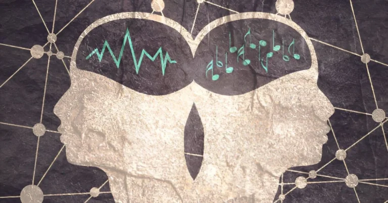
Near-death experiences through the lens of neuroscience
At the boundary between life and death, the brain does not shut down all at once.
It may instead produce a final episode of coordinated activity underlying powerful subjective experiences.
When the heart suddenly stops beating, intuition suggests that consciousness should vanish instantly. Neuroscience research over the past decade, however, tells a very different story. Biological death is not a switch that flips off. It is a transition. For several seconds, sometimes up to tens of seconds, brain networks oscillate between collapse and bursts of intense activity, generating mental states of unusual vividness.
This insight emerged from a now landmark study. In 2013, researchers at the University of Michigan recorded the brain activity of rats subjected to experimentally induced cardiac arrest. They expected an immediate drop in neural signals. Instead, they observed a surge in gamma waves, fast oscillations typically associated with attention, conscious perception, and multisensory integration. Remarkably, this activity was even more organized and synchronized than during wakefulness.
These findings prompted scientists to examine the final moments of human brain activity in intensive care settings. In 2023, recordings from patients experiencing cardiac arrest revealed similar patterns. Just before total electrical collapse, frontoparietal networks showed transient signs of reactivation. Even deprived of oxygen, the brain appeared to mount a final effort to preserve a minimal level of integration, a last surge of organization against disintegration.
This fragile interval, neither fully alive nor yet devoid of activity, is where many near-death experiences take shape. It is within this in-between state that perceptions of light, sensations of floating, and feelings of extraordinary clarity emerge. Not after the brain has died, but precisely while it is still struggling to sustain a fragment of consciousness.
🔗Read also: The conscious brain: Inside a scientific showdown
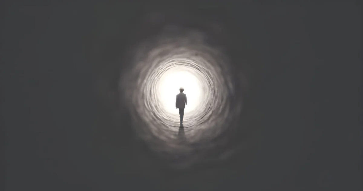
Light, tunnels, and out-of-body experiences
Accounts of near-death experiences display a striking consistency across individuals. People frequently describe an intense light, the sensation of moving through a tunnel, an irresistible pull toward a luminous point, or the impression of floating above their own body, as though consciousness had temporarily detached from physical form. These experiences are neither random nor chaotic. They reflect how specific brain regions respond when oxygen supply becomes critically reduced, revealing shared neurobiological mechanisms across testimonies.
The luminous tunnel, for instance, is one of the best-understood features. The visual cortex is particularly vulnerable to hypoxia. Peripheral visual areas fail before central regions, mechanically narrowing the visual field. Studies of induced loss of consciousness, notably in fighter pilots, show that individuals often report a central light surrounded by encroaching darkness. The attraction toward light commonly described in near-death experiences aligns precisely with this physiological process, mirroring the brain’s predictable response when its operating conditions are profoundly altered.
Out-of-body experiences strike at the core of bodily identity. The temporoparietal junction, located at the intersection of the temporal and parietal lobes, plays a crucial role in generating the sense that “I am located within my body.” This region continuously integrates visual, tactile, vestibular, and proprioceptive signals to maintain a coherent representation of the body in space. When its functioning is disrupted, this representation can fragment, leading to a loss of bodily anchoring and, in some individuals, the subjective impression that the point of conscious perspective has shifted outside the body.
This phenomenon was first observed clinically in the early 2000s by neurologist Olaf Blanke and his team. During epilepsy surgery preparation, a fully conscious patient repeatedly reported out-of-body sensations when physicians electrically stimulated a highly specific area of the right temporoparietal junction. She described floating above her body and observing herself from an external vantage point. As soon as stimulation stopped, the experience vanished instantly. The effect was reproducible, anatomically precise, and strictly dependent on a defined cortical region, ruling out imagination or dream-like explanations.
Subsequent experiments replicated similar illusions in healthy participants. Virtual reality studies demonstrated that artificially inducing sensory conflicts can shift the sense of bodily self-location. By synchronizing tactile stimulation with the visual perspective of a body filmed from behind, researchers were able to displace where individuals felt “located” in space. This mismatch alone was sufficient to convince the brain that the center of experience had moved.
These findings illuminate what occurs during near-death experiences. In this context, hypoxia profoundly disrupts sensory processing. Signals that normally stabilize embodiment, arising from vision, vestibular input, and proprioception, become unstable and incoherent. Faced with this extreme disturbance, the brain does what it always does: it attempts to reconstruct a unified experience from fragmented information. A decentered perspective may then emerge, offering a subjective viewpoint from which the scene regains coherence. This is not consciousness leaving the body, but a transient reconfiguration of neural representations of the body and self under severely altered physiological conditions.
🔗Explore further: Altered states of consciousness: Inside the sacred rotation of the Dervishes
Conscious experience in a brain under extreme stress
Near-death experiences do not arise from an inactive brain. They emerge from a brain that is still functioning, albeit under extreme physiological stress. Clinical data converge on a crucial point: once neural activity truly ceases, no experiential content can be encoded into memory. Near-death narratives therefore correspond to a fleeting window during which brain activity is profoundly altered but remains sufficient to generate conscious experience and allow it to be later remembered.
The psychological impact of these episodes is profound. Many individuals report lasting transformations, including reduced fear of death, shifts in life priorities, and a renewed sense of meaning. These effects do not contradict their cerebral origins. On the contrary, the combination of intense emotion, physiological struggle, and sensory disruption strongly enhances memory consolidation. The experience becomes vivid, foundational, and enduring.
Ultimately, science reveals the remarkable power of a brain that, even as its foundations crumble, strives to preserve narrative coherence and a final image of the world. Near-death experiences do not tell us what lies beyond. They reveal what remains active in the seconds as life recedes. A brain that resists, until its very last electrical impulse.
References
Blanke, O., Ortigue, S., Landis, T., & Seeck, M. (2002). Stimulating illusory own‐body perceptions. Nature, 419(6904), 269–270.
Borjigin, J., Lee, U., Liu, T., Pal, D., Huff, S., Klarr, D., … & Mashour, G. A. (2013). Surge of neurophysiological coherence and connectivity in the dying brain. Proceedings of the National Academy of Sciences, 110(35), 14432–14437.
Ehrsson, H. H. (2007). The experimental induction of out-of-body experiences. Science, 317(5841), 1048.
Lempert, T., Bauer, M., & Schmidt, D. (1994). Syncope: A videometric analysis of 56 episodes of transient cerebral hypoxia. Annals of Neurology, 36(2), 233–237.
Marti, S., King, J.-R., & Dehaene, S. (2015). Time-resolved decoding of two processing chains during dual-task interference. Neuron, 88(6), 1297–1307.
Mobbs, D., & Watt, C. (2011). There’s nothing paranormal about near-death experiences: How neuroscience can explain seeing bright lights, meeting the dead, or being convinced you are one of them. Trends in Cognitive Sciences, 15(10), 447–449.
Parnia, S., Spearpoint, K., & de Vos, G. (2014). AWARE—AWAreness during REsuscitation—A prospective study. Resuscitation, 85(12), 1799–1805.
Vicente, R., et al. (2023). Enhanced frontal and parietal EEG activity in the dying human brain. Resuscitation, 188, 109–116.
Sara Lakehayli
PhD, Clinical Neuroscience & Mental Health
Associate member of the Laboratory for Nervous System Diseases, Neurosensory Disorders, and Disability, Faculty of Medicine and Pharmacy of Casablanca
Professor, Higher School of Psychology