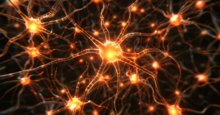
How Astrocytes Reshape the Brain
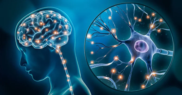
For a long time, we believed that neurons alone were responsible for shaping our thoughts, memories, and adaptive capabilities. This perspective, firmly rooted in classic neuroscience research, led to the significant undervaluation of glial cells, particularly astrocytes. For decades, these cells were merely regarded as structural support, a biological scaffold meant to hold neurons in place and provide them with nutrients. However, modern science now paints a very different picture: far from being passive, astrocytes stand out as key contributors to cerebral plasticity and neuronal repair.
In neurological disorders, a sudden event or lesion disrupts the brain’s usual balance, impairing cognitive functions. Nevertheless, the brain’s ability to adapt and heal itself depends largely on the astrocyte response to such injuries. Contrary to the enduring belief that the brain is rigidly fixed in its organization, recent findings show these cells play a pivotal role in modulating cerebral plasticity, particularly after lesions that compromise the blood-brain barrier. A recent study published in Nature Medicine explored the regenerative functions of astrocytes, revealing the molecular mechanisms that enable these cells to drive the repair of brain tissue.
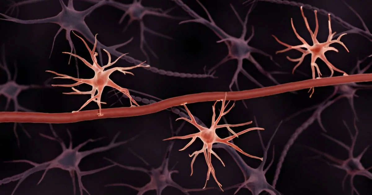
Glial cells in action: the shift from structural support to tissue repair
This research is particularly important because it challenges our long-held understanding of astrocytes in cerebral plasticity and opens the door to new therapeutic possibilities. In conditions affecting the brain, such as strokes, head injuries, or certain neurodegenerative diseases, the reaction of astrocytes is critical. Their activity governs both the formation of a glial scar and the tissue’s regenerative capacity.
🔗 Read also: Timeless regeneration: Uncovering the brain’s stem cell secrets
A team led by Swetlana Sirko and Magdalena Götz at Ludwig Maximilian University of Munich addressed this question by examining cerebral cortex samples taken from patients with cerebral cavernous malformations, a condition caused by abnormal blood vessels in the brain. Because these hemorrhagic lesions abruptly breach the blood-brain barrier, they provide an ideal model for studying how astrocytes respond to an altered microenvironment.
The researchers found that astrocytes in damaged areas proliferated more than those in healthy tissue. A particularly noteworthy marker emerged: GALECTIN3, a protein linked to proliferating reactive astrocytes. This biomarker appears to be specific to regions where the blood-brain barrier is compromised. Furthermore, these cells do more than simply multiply: they acquire properties similar to neural stem cells, potentially contributing to neuronal regeneration. This finding transforms our understanding of glial plasticity and opens promising therapeutic avenues.
these cells do more than simply multiply: they acquire properties similar to neural stem cells, potentially contributing to neuronal regeneration.
The team employed a range of advanced techniques, immunohistochemistry, mass spectrometry, and in vitro cell cultures, to conduct the study. One key element was the analysis of the patients’ cerebrospinal fluid. Bathing the central nervous system, this fluid contains numerous regulatory signals. By comparing samples from patients with cerebral cavernous malformations to those from patients with meningiomas (benign tumors without vascular rupture), the researchers identified a critical regulatory component: LGALS3BP, a protein that interacts with GALECTIN3 and stimulates astrocyte proliferation.
🔗 Explore further: From loss to repair: Stem cells offer new hope for Parkinson’s
Shaping astrocyte plasticity for healing
The involvement of LGALS3BP in astrocyte plasticity is vital. More than just a biomarker, it plays a functional role by shaping the behavior of reactive astrocytes. Its inhibition, tested in vitro, significantly diminishes astrocyte proliferation and neurosphere formation, structures that showcase the stem cell potential of these cells in culture. This result offers promising possibilities for controlling glial scar formation and enhancing functional recovery after brain trauma.
These discoveries have far-reaching implications beyond hemorrhagic injuries. In neurodegenerative conditions, the glial response plays a complex role. It acts as a first line of defense against damage, but it can also aggravate lesions by creating an environment that hinders neurogenesis. Manipulating this response has therefore become a key therapeutic goal, not only for traumatic injuries but also for conditions such as Alzheimer’s disease, Parkinson’s disease, and post-traumatic epilepsy.
🔗 Discover more: Kafka’s Metamorphosis and the voice of silence
While this study provides inspiring insights, it is just the beginning of our quest to understand how astrocytes help repair the brain. The main challenge will be to determine how these mechanisms function in vivo, within a living, active brain. Gaining a more thorough grasp of the interplay among GALECTIN3, LGALS3BP, and other factors that shape glial plasticity may pave the way for customized recovery strategies tailored to each disorder. In this sense, today’s research merely scratches the surface of a field with immense therapeutic potential, redefining our view of the brain as a constantly evolving system.
References
Sirko, S., Schichor, C., Della Vecchia, P., Metzger, F., Sonsalla, G., Simon, T., Bürkle, M., Kalpazidou, S., Ninkovic, J., Masserdotti, G., Sauniere, J.-F., Iacobelli, V., Iacobelli, S., Delbridge, C., Hauck, S. M., Tonn, J.-C., & Götz, M. (2023). Injury-specific factors in the cerebrospinal fluid regulate astrocyte plasticity in the human brain. Nature Medicine, 29, 3149–3161.