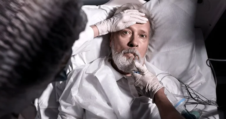
When silence speaks: The enigma of Mr Tan
Some silences speak louder than words. In Mr Tan’s case, his silence was not an absence, but a riddle. When he arrived at Dr. Paul Broca’s office, he brought with him a fundamental question: How does the brain create what makes us human, our language, our ability to name the world around us, to share our thoughts and emotions? And yet, Mr Tan had only one word to give: tan. This single sound, repeated over and over, became the key to a revolutionary discovery. His case would open a door long sealed, shedding light on the connection between our cognitive functions and the underlying brain networks that support them.
Mr Tan’s story is not merely one of loss, but also one of revelation. It shows how an apparent malfunction in brain function can illuminate hidden mechanisms. His silence prompts us to reconsider what it means to “speak” or “express,” and to marvel at the fascinating mysteries of human biology. Blending science, philosophy, and the weight of individual destinies, Mr Tan’s story reminds us that even in loss, the brain finds ways to share its secrets.
Piecing together the mind: Broca’s landmark discovery
When Mr Tan stepped into Dr. Broca’s office, his silence made a far more striking impression than any words might have. However, this silence was not a void. It was charged with meaning, like an ancient text whose richness is only suspected, yet remains indecipherable. Mr Tan spoke only one word: “tan.” At every question, every attempt at conversation, this simple sound returned, stubborn and puzzling, like a single note replayed on a broken instrument. His body, meanwhile, presented its own mystery, an open book written in a language no one could decipher.
This patient, destined to become one of the most famous figures in neuroscience, carried within him a truth no one yet suspected. Dr. Broca, fascinated by the puzzle, perceived a key to unlocking one of the brain’s great secrets in that silence. After Mr Tan’s death, the autopsy revealed a lesion in a specific region of the left hemisphere, now known as Broca’s area. This discovery was a turning point: for the first time, a direct link was established between a particular brain region and a specific function, namely language production. Where the body seemed disordered, a subtle design emerged, an architecture of thought woven into physical matter.
After Mr Tan’s death, the autopsy revealed a lesion in a specific region of the left hemisphere, now known as Broca’s area.
Mr Tan’s brain was like a mosaic. Some tiles remained intact, allowing his thoughts to flourish in silence, while others were fractured, trapping his voice. His comprehension stayed sharp, and his intellect remained bright, but words were lost in the echo of that small lesion. Still, this silence did not appear to crush him. He carried a quiet dignity, almost serene, as if his body and brain had made their peace with the loss.
Dr. Broca did not merely see a patient in Mr Tan. He saw a gateway to a broader truth: the idea of a fragmented yet organized brain, where each region fulfills a distinct function. In his strange mutism, Mr Tan was more than a patient; he was an entry point into the inner workings of the brain. This emblematic case not only revealed the vulnerabilities of speech, but also the resilience of the human spirit when faced with the brain’s hidden tempests.
Although seemingly mute, Mr Tan was not devoid of thought. Contrary to the intuitive notion of a complete absence, his condition revealed a break, a subtle disconnection. His thoughts were vivid, coherent, and fully formed, but the circuits enabling them to become language were severed. Like a river whose course is blocked by a dam, his ideas were kept captive, unable to flow out into words. This image of a trapped mind highlights a fundamental truth about our brain: rather than functioning as a machine made of separate parts, it acts as a complex network, an interwoven system in which each region collaborates on specific tasks.
Cognitive abilities do not emerge from an isolated region; they arise from the harmony of interconnected circuits. When even one link in this chain breaks, the function it supports falters, sometimes irreversibly. For Mr Tan, the lesion to Broca’s area caused a permanent loss of speech production. Still, this loss did not negate who he was. His intellect remained intact, and he understood the world around him. He responded with gestures, facial expressions, and that single word, “tan”, which served, however limited, as an attempt to exist in an imposed silence. Although his brain could not reconstruct those lost circuits, it continued to function in other domains, sustaining an inner life that, though unheard, was profoundly human.
Cognitive abilities do not emerge from an isolated region; they arise from the harmony of interconnected circuits. When even one link in this chain breaks, the function it supports falters, sometimes irreversibly.
Aphasia and beyond: tracing the fragile roads of speech
Language, one of the most extraordinary abilities of the human brain, allows us to transform thoughts into articulated sounds, build bridges among individuals, and breathe life into the most abstract of ideas. Beneath this apparent ease lies a complex mechanism, an infinitely precise interplay among different brain areas. Aphasia, the term for the loss or alteration of language, arises when this harmony is disrupted. It can affect word production, comprehension, or even the ability to read and write, revealing just how interdependent these processes really are.
In the brain, language does not reside in one isolated region, but rather in a collaboration among several key areas. Broca’s area, in the left hemisphere, plays a central role in producing spoken language. It directs the movements needed to form sounds and organizes them into words and sentences. Meanwhile, Wernicke’s area, also in the left hemisphere, creates meaning from what we hear or plan to say. These regions, though distinct, communicate through networks of nerve fibers, such as the arcuate fasciculus, forming an integrated system in which each component is indispensable.
In the brain, language does not reside in one isolated region, but rather in a collaboration among several key areas.
When a part of this network is damaged, as in certain types of aphasia, speech can become fragmented or reduced to scraps, revealing the fragile nature of this faculty we often take for granted. Aphasias vary according to the areas affected: a lesion in Broca’s area (Broca’s aphasia), as in Mr Tan’s case, causes speech difficulties, whereas damage to Wernicke’s area (Wernicke’s aphasia) can interfere with comprehension, turning speech into a cascade of disordered, meaningless words. These disruptions are not simply symptoms; they are windows into the deeper processes of language and the ways the brain links thought, sound, and meaning.
Recent advances in neuroscience have greatly expanded our understanding of language mechanisms and recovery processes after brain injury, largely thanks to neuroplasticity. This capacity for the brain to reorganize its networks is remarkably evident in aphasic patients, as demonstrated in a landmark 1983 study by J. Cambier. This research revealed not only the boundaries but also the possibilities of functional compensation following severe hemispheric injury.
In the study, two right-handed patients with right-sided hemiplegia and aphasia, both caused by a left-hemisphere stroke, were observed. The most striking case involved a patient who had lost nearly all of her language abilities. Through an extended rehabilitation program, she gradually recovered certain linguistic skills, including oral and written expression and comprehension, though her ability to build complex sentences remained limited. This partial recovery demonstrated that her right hemisphere, typically less involved in language, had taken over some linguistic functions due to the combined effects of the left-hemisphere lesion and intensive therapeutic stimulation. While this compensation remained limited, it nonetheless represented a major reorganization of the brain, revealing its remarkable resilience in the face of adversity.
Her story took a dramatic turn two years later when she suffered a second stroke, this time in the right hemisphere. She then lost all linguistic abilities, confirming that although the right hemisphere can partially compensate for language function, it is less efficient and cannot fully replace the dominant hemisphere. At autopsy, bilateral infarcts in the sylvian territories were found, underscoring the scope of the damage and reminding us that the adult brain’s plasticity has its limits.
However, it is in the developing brain that plasticity reaches its peak, as illustrated by the heartrending case of Matthew, recounted by researcher David Eagleman. Matthew was only three years old when he was diagnosed with Rasmussen’s encephalitis, a rare disease that triggers unbearable seizures. The only option to save him was a hemispherectomy, removing an entire hemisphere of his brain. In the wake of this radical procedure, Matthew was left unable to speak, walk, or perform even the simplest tasks. His devastated parents faced an uncertain future, fearing the irreversible consequences of such an intervention.
Still, Matthew’s young, adaptable brain embarked on a remarkable reorganization. The remaining hemisphere harnessed every resource at its disposal, step by step assuming the functions that had been lost. Three months after the surgery, Matthew began to speak again, progressing through the stages typical of a toddler learning language for the first time. This phenomenon, arising from a rewiring of neural circuits and intensified use of intact regions, demonstrates the unique capacity of the developing brain to compensate for massive loss.
Today, Matthew leads a functional life. Observers unfamiliar with his past would scarcely notice his slight limp or mild weakness in his right hand. His story highlights not only the exceptional power of the young brain’s plasticity, but also the crucial importance of early intervention and targeted rehabilitation in harnessing its ability to adapt.
These clinical cases underscore several essential points. First, patient age is pivotal: in younger individuals, brain plasticity is generally more dynamic, allowing for broader functional reorganization. In adults, as shown in Cambier’s study, compensatory abilities are still possible but less robust. Second, post-injury plasticity involves complex processes, such as functional reorganization, the activation of alternative neural pathways, and the recruitment of often underutilized cortical circuits. Finally, the intensity and duration of rehabilitation prove vital in maximizing the brain’s adaptive potential.
How brain injuries expose human resilience
The stories of Mr Tan, Matthew, and countless other patients confronting profound cerebral loss form more than just a series of clinical cases. Together, they demonstrate the complexity of our brain in action. Far from being an empty void, Mr Tan’s silence mirrored a still-intact process deep within his mind. He thought, understood, and lived. What he had lost was not his inner life, but one way of expressing it. Matthew, on the other hand, showed us that after a hemispherectomy, seemingly a life-altering blow, the developing brain can exhibit astonishing plasticity. With intensive rehabilitation, his remaining hemisphere took over essential functions, enabling him to relearn speech and to resume a fulfilling life.
As different as these accounts may be, they converge around the reality that our brain is not a rigid assembly of functions but rather a living, dynamic tapestry capable of reinventing itself in the face of extreme obstacles. Through his study of Mr Tan, Paul Broca pioneered our understanding of how specific brain regions give rise to fundamental capacities like language. Since then, neuroscientists have greatly expanded that knowledge, showing that reorganization can even occur after radical procedures such as hemispherectomy or massive functional losses.
These clinical narratives, blending scientific insight with human stories, remind us how complex, and profoundly resilient, the brain can be. Anything we regard as certain, sturdy, and permanent can vanish in an instant. Nevertheless, this fragility is also a form of strength. Even in adversity, humanity possesses a remarkable capacity to adapt, to transform loss into new potential, and to forge new pathways.
References
Cambier, J., Elghozi, J. L., Signoret, J. L., & Henin, D. (1983). Contribution de l’hémisphère droit au langage des aphasiques : Disparition de ce langage après lésion droite. Revue neurologique, 139(1), 55–64.
Eagleman, D. (2020). Livewired: The inside story of the ever-changing brain. Pantheon Books.

Sara Lakehayli
PhD, Clinical Neuroscience & Mental Health
Associate member of the Laboratory for Nervous System Diseases, Neurosensory Disorders, and Disability, Faculty of Medicine and Pharmacy of Casablanca
Professor, Higher School of Psychology


